Signals that help move the left hand largely originate in the right motor cortex. Information about touch on that hand also reaches the opposite cerebral hemisphere. This familiar arrangement raises an evolutionary puzzle: why do so many important nerve pathways cross from one side of the nervous system to the other?
University of Minnesota neuroscientists Martin W. Wessendorf and Daniel Delie Wang propose that the answer may begin with ancient visual circuits. Their paper in Frontiers in Neuroanatomy reviews existing anatomical research and develops an evolutionary hypothesis. It does not report an experiment demonstrating how crossed wiring originated.
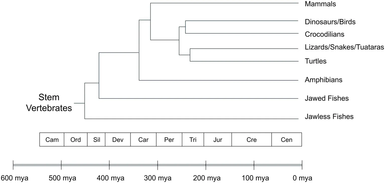
The researchers focus on the optic tectum, a midbrain structure called the superior colliculus in mammals. This structure helps animals orient toward or respond to events around them. Its broad distribution across living vertebrates suggests that it existed in their distant common ancestor. Modern jawless fishes, including lampreys, provide part of the evidence behind that inference.
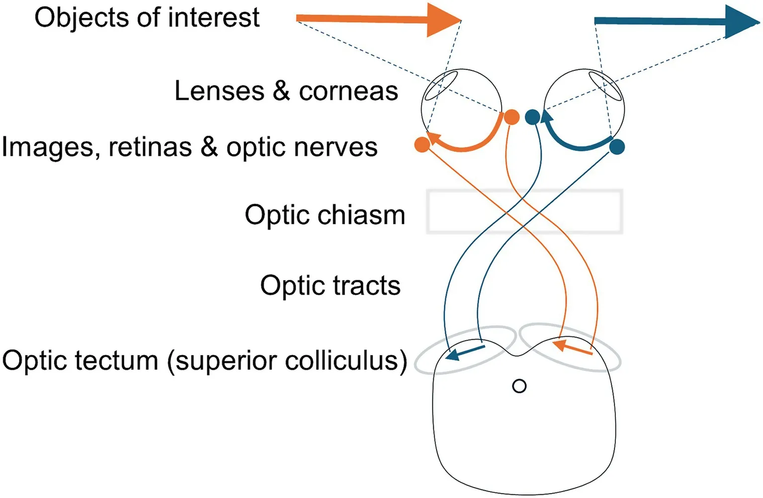
Visual information reaches it through pathways organized around the opposite side of visual space. In humans, information from the right visual field reaches the left side of the brain, and vice versa. This arrangement concerns visual fields, not a simple rule assigning each entire eye to the opposite hemisphere.
Each eye’s lens reverses the image that reaches the retina. At the optic chiasm, nerve fibers are arranged so that information about the same side of visual space travels together. Because both human eyes see parts of both visual fields, some fibers cross there while others remain on the same side.
The optic tectum contains an organized map of visual space. Deeper layers receive other sensory information, with spatial maps that roughly align with the visual map. Still deeper circuits help generate movements, allowing the structure to connect information about surrounding events with appropriate responses.
Wessendorf and Wang propose that this existing arrangement influenced pathways that evolved later. Adding touch, hearing and movement circuits around an established opposite-side map may have required fewer changes than building a different arrangement. This is the central hypothesis, rather than an observed reconstruction of ancient brains.
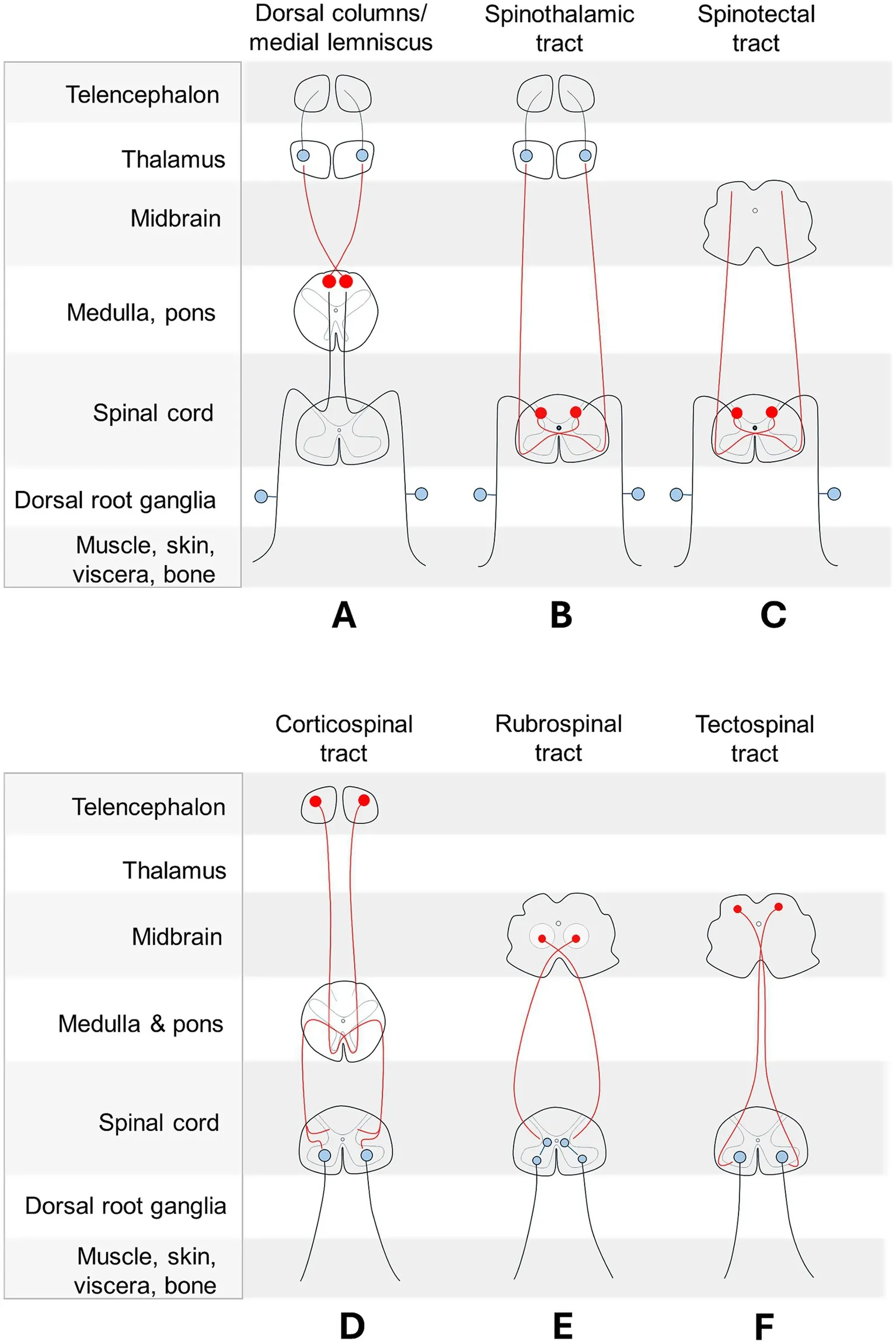
The authors examined how bodily sensations reach the brain across vertebrate groups. One major route, the dorsal column pathway, carries information about light touch, vibration and body position. Signals initially ascend the spinal cord on the same side as their source.
After reaching the lower brainstem, they connect with neurons whose fibers cross the midline. The information then continues toward the thalamus and, in mammals, the cerebral cortex. The familiar crossed relay has been described in amphibians, reptiles, birds and mammals, supporting an inferred origin in their shared ancestry.
Another route, the spinothalamic pathway, carries information including pain, temperature, itch and some touch. Its fibers cross within the spinal cord before ascending toward the thalamus. Thus, pathways can deliver information to the opposite side of the brain while crossing at different levels of the nervous system.
Several descending pathways also cross the midline. The tectospinal pathway carries signals from the optic tectum toward the spinal cord and contributes to orienting movements. The rubrospinal pathway begins in the midbrain’s red nucleus and participates in movement control, including limb control in many animals.
These pathways vary across vertebrate groups, so the paper does not describe one identical circuit plan in every species. Their distribution nevertheless suggests that several crossed systems existed before mammals developed their characteristic cerebral cortex. The authors argue that this older organization helped constrain the connections added later.
The corticospinal pathway presents a particularly interesting example. It connects the mammalian cerebral cortex with the spinal cord and supports precise movements in humans. In many other mammals, its connections are especially prominent in spinal regions that process sensory information.
Wessendorf and Wang propose that fine motor control recruited a pathway with an ancestral role in regulating bodily sensation. Under that interpretation, its crossed organization could have accompanied the change in function. The proposal concerns evolutionary reuse, rather than evidence that the pathway performs only one task in each species.
The paper distinguishes opposite-side connections from lateralization, the uneven distribution of functions between the brain’s two sides. Handedness is an example of lateralization. Crossed wiring, by contrast, can be broadly symmetrical: each hemisphere connects with the opposite side of the body.
The researchers reconstructed possible histories by comparing traits reported in related animal groups. A feature shared widely across a group may have existed in its last common ancestor. This approach provides evolutionary inferences, but incomplete sampling and changes within individual lineages complicate the reconstruction.
Crossing is also far from universal. Some pathways project to both sides, while others remain largely on the same side. The cerebellum generally coordinates movement on its own side of the body, sometimes through connections that cross twice.
The visual explanation also has historical roots. Santiago Ramón y Cajal proposed in 1898 that visual organization might have influenced other crossed systems. The new paper develops that reasoning around the optic tectum and the sequence in which different pathways appear to have evolved.
Development introduces another distinction between mechanism and history. Molecular signals help guide growing nerve fibers toward, across or away from the midline as a nervous system forms. Identifying those signals explains how a connection develops in an individual, but does not automatically establish why that arrangement became widespread during evolution.
Other hypotheses invoke an ancestral body-axis twist, developmental scaffolds or potential advantages in responding to injury. The authors discuss difficulties with these explanations rather than experimentally eliminating them. Their own account likewise needs tests that could distinguish it from alternatives.
The authors suggest examining Cambrian vertebrate fossils for evidence of an optic chiasm and optic tectum. Their hypothesis predicts that both structures existed in the common ancestor of living vertebrates. Exceptionally preserved remains or detailed reconstructions of skull interiors might help, although ordinary fossils cannot reveal individual nerve connections.
Living animals offer additional tests. The paper predicts that crossed pathways connecting the forebrain with the spinal cord will prove widespread among birds. It also predicts comparable connections in crocodilians, a group the authors identify as insufficiently examined for this pathway.
A further prediction concerns wiring costs, including the resources needed to maintain connections and transmit signals. The authors argue that preserving an established crossed arrangement may have been more economical than reorganizing multiple systems. They did not perform a new comparison demonstrating those savings, so the efficiency claim remains a hypothesis.
The proposal shifts attention toward how inherited structures constrain later evolution. It does not establish why the earliest visual pathways crossed in the first place. The next challenge is to determine whether fossil evidence, broader anatomical surveys and explicit wiring comparisons support the optic tectum’s proposed role in shaping the circuits that followed.
These resources combine recent research on sensory integration and nerve-fiber guidance with foundational work on brain organization.
Functional specialisation of multisensory temporal integration in the mouse superior colliculus
Recordings from more than 5,000 neurons in awake mice reveal how this midbrain structure combines visual and auditory timing information. The findings illuminate its present-day function without establishing why crossed pathways evolved. (Nature Communications, October 2025)
Corticospinal Tract Development, Evolution, and Skilled Movements
This recent review connects differences in corticospinal development across species with skilled movement and manual dexterity, updating the background on a central pathway discussed in the story. (Movement Disorders, April 2025)
Plexin B3 guides axons to cross the midline in vivo
Experiments in zebrafish embryos identify a molecular contributor to retinal nerve fibers crossing at the optic chiasm. The study addresses how crossed wiring develops, rather than its evolutionary origin. (Frontiers in Cellular Neuroscience, April 2024)
Research findings are available online in the journal Frontiers in Neuroanatomy.
The original story “Why does the right side of our brain control the left side of our body? New research may have the answer” is published in The Brighter Side of News.
Like these kind of feel good stories? Get The Brighter Side of News’ newsletter.
The post Why does the right side of our brain control the left side of our body? New research may have the answer appeared first on The Brighter Side of News.


Leave a comment
You must be logged in to post a comment.