A notebook or small box on a table became easier to detect for some people whose retinal disease had severely damaged their sight. An experimental treatment gave surviving retinal nerve cells a new ability to respond to light. Special goggles supplied the signals those cells needed to convey limited visual information.
The ten-person PIONEER trial, reported in The New England Journal of Medicine, primarily assessed safety. Seven participants showed increased light sensitivity after treatment, and four showed consistent gains across several behavioral tests. Improvements varied considerably, and the approach did not restore normal or high-resolution vision.
José-Alain Sahel of the University of Pittsburgh and Botond Roska of the Institute of Molecular and Clinical Ophthalmology Basel and University of Basel led the work. GenSight Biologics sponsored the study and managed its data. The trial included the participant whose partial visual recovery the team first reported in 2021.
Retinitis pigmentosa encompasses inherited diseases that progressively destroy photoreceptors, the retina’s natural light-sensing cells. Changes in more than 100 genes can cause the condition. Treatments aimed at replacing a faulty gene therefore apply to particular genetic forms and generally require suitable surviving cells.
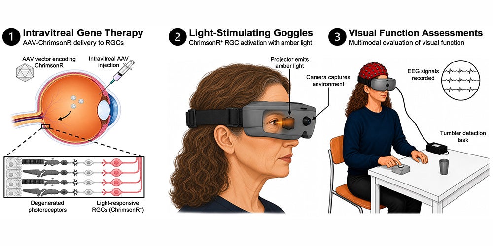
Optogenetics takes a different route by introducing instructions for a light-sensitive protein into neurons that remain. The PIONEER treatment, called GS030, targets retinal ganglion cells, which normally transmit processed visual signals toward the brain. It seeks to bypass lost photoreceptors rather than replace them or repair the original mutation.
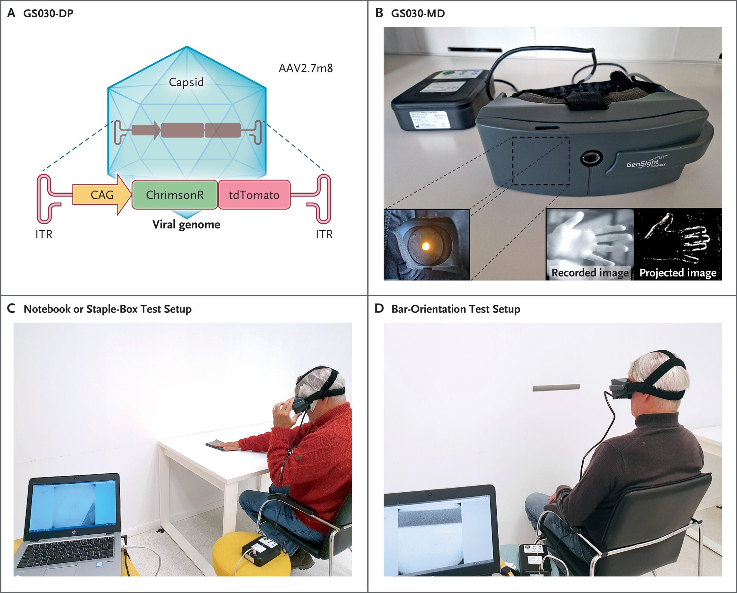
Participants received a single injection into their worse-seeing eye. An adeno-associated viral vector carried instructions for ChrimsonR, a protein activated by amber light. The researchers tested three increasing doses, with three participants in each of the lower groups and four in the highest.
Camera-equipped goggles detected changes in the surrounding scene and projected corresponding amber pulses onto the retina. Those pulses activated the modified cells, supplying a new source of visual signals. The combination was designed to work independently of the disease-causing mutation, although this small study could not establish whether genetic subtypes respond differently.
The participants were six women and four men, averaging approximately 55 years old at treatment. Their worse-seeing eyes initially ranged from hand-motion perception to light perception or no light perception. All ten underwent safety assessments and light-sensitivity testing, while eight completed the principal object and orientation tests.
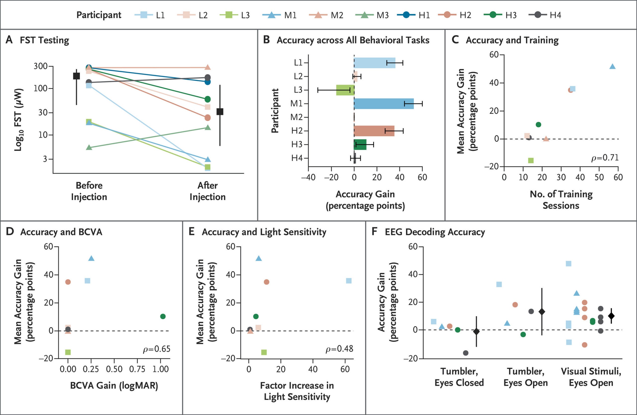
With the goggles, seven participants became more sensitive to light after the injection. Six met a threshold corresponding to approximately a fourfold improvement in sensitivity. That threshold came from published evidence but was not prespecified in the trial protocol, an important qualification when interpreting an exploratory result.
Behavioral testing compared performance with the goggles switched on and off, while the untreated eye remained patched. Tasks included detecting, locating and touching a notebook or staple box, and identifying a bar’s orientation. Four of eight participants showed consistent positive changes across tests, according to a response definition that also was not prespecified.
Those four participants maintained improvements in notebook detection across repeated sessions spanning roughly one to one-and-a-half years. Some described an unfamiliar vibrating sensation when perceiving objects. However, group-average estimates were imprecise, with confidence intervals spanning no improvement for several tasks, so the results do not establish a reliable benefit for every patient.
Researchers recorded 34 eye-related adverse events among nine participants. Twenty-three were mild and ten moderate. One event was classified as severe: a temporary central retinal artery blockage immediately after injection, which resolved within minutes after treatment.
Inflammation inside the eye occurred in seven participants and was more common in the highest-dose group. These episodes resolved with standard treatment without lasting consequences. Retinal imaging generally remained stable, providing reassurance within the limits of the small cohort and available follow-up.
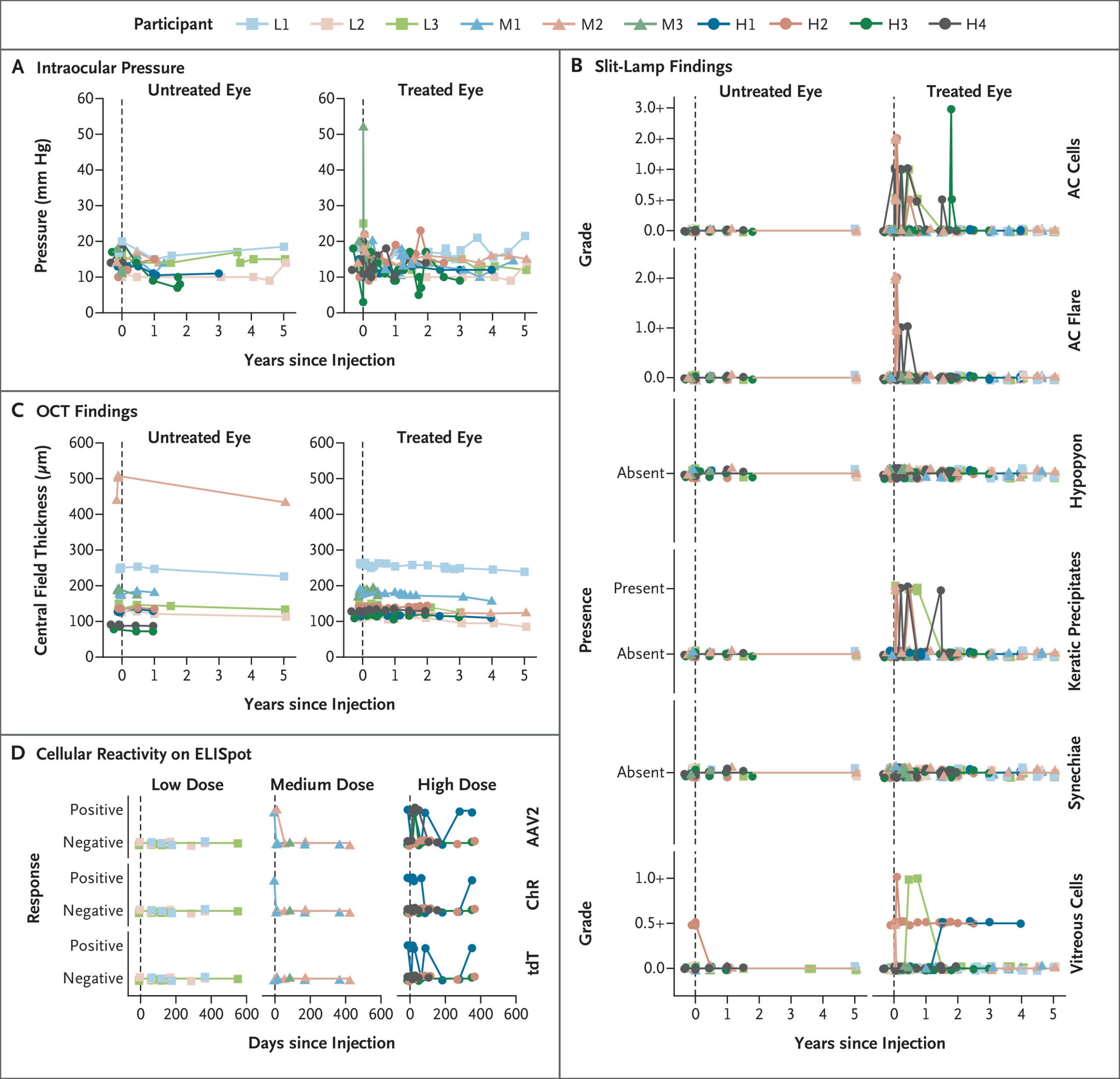
Investigators did not attribute any systemic adverse event to the study drug or injection procedure. Three mild systemic events were attributed to the goggles. No participant stopped testing because of an adverse event, although other reasons prevented some participants from completing every assessment.
At the November 2024 data cutoff, four participants had completed five years of follow-up. Several others had been followed for two to four-and-a-half years, while two withdrew earlier. These observations support further evaluation, but ten participants cannot establish the frequency of uncommon complications or definitively settle long-term safety.
Participants trained with the goggles after receiving the injection, learning to use a form of perception unlike ordinary sight. Those classified as responders averaged 36.5 training sessions, compared with 15.3 among nonresponders. Training sessions were separate from the formal behavioral tests.
Greater training exposure was associated with better performance, suggesting rehabilitation may matter. However, the correlation estimate was uncertain, and its confidence interval included no association. The study did not randomly assign training intensity, so it cannot show that additional sessions caused the better outcomes.
Brain recordings provided another exploratory check. Researchers tested five participants, selecting four because they had already shown behavioral responses. Some signals were consistent with visual information reaching the cortex, but several estimates remained imprecise and did not clearly exclude no effect.
The trial was open-label and nonrandomized, and all visual-function assessments were exploratory. Some procedures were developed after the first participant reported responses and were then standardized. These features make larger, controlled studies important for separating treatment effects from training, variability and other influences.
Several biological constraints limit the current system’s resolution. The treated ganglion cells occupy a ring around the fovea rather than recreating the retina’s normal spatial organization. Direct activation also bypasses natural retinal processing and does not reproduce the different response patterns of normal ganglion-cell types.
A single light sensor cannot restore color vision, and advanced disease leaves relatively few ganglion cells available. Future refinements could include eye tracking, image preprocessing tailored to the treated retinal region and improved stimulation methods. These remain proposals rather than demonstrated solutions in this trial.
“Our next major challenge is to achieve high-resolution vision that would allow patients to recognize faces,” Roska said. For now, the evidence supports a narrower achievement: some people with advanced retinal degeneration gained modest visual capabilities using surviving cells and specialized equipment. Establishing how consistently those gains translate into everyday independence will require further research.
These resources trace the approach’s development and explain the broader biology of inherited vision loss.
Partial recovery of visual function in a blind patient after optogenetic therapy: Reports the original single-participant demonstration that preceded the expanded PIONEER results. (Nature Medicine, 2021)
Optogenetic therapy: high spatiotemporal resolution and pattern discrimination compatible with vision restoration in non-human primates: Presents preclinical work on the vector and light-sensitive protein used in the human approach. (Communications Biology, 2021)
Independent optical excitation of distinct neural populations: Describes light-sensitive tools including Chrimson, foundational to later optogenetic applications. (Nature Methods, 2014)
Optogenetic restoration of retinal ganglion cell activity in the living primate: Examines light-evoked activity in modified retinal neurons, helping connect cellular responses with therapeutic development. (Nature Communications, 2020)
Retinitis Pigmentosa: Explains symptoms, inheritance, diagnostic testing and rehabilitation for people with the condition. (National Eye Institute, 2025)
Research findings are available online in The New England Journal of Medicine.
The original story “Experimental treatment helps some blind patients see objects” is published in The Brighter Side of News.
Like these kind of feel good stories? Get The Brighter Side of News’ newsletter.
The post Experimental treatment helps some blind patients see objects appeared first on The Brighter Side of News.


Leave a comment
You must be logged in to post a comment.