A repair cell’s movement can do more than carry it into damaged tissue. An experimental material uses that movement to release healing proteins, turning a tiny mechanical force into a precisely placed biological signal.
The dressing captures selected growth factors, holds them inactive and releases them when cells pull on it. Tests found improved blood vessel formation in rat bone injuries, smaller mouse wounds and greater cell migration in human skin maintained outside the body.
The Imperial College London study, published in Nature Materials, extends an earlier laboratory concept into repairing tissues. Lead author Magdalene Ho and senior author Benjamin Almquist developed the approach with colleagues studying biomaterials and tissue repair.
These are preclinical results, rather than evidence from treated patients. The human experiments used surgical skin samples kept alive in the laboratory, allowing researchers to examine how tissue interacted with the material.
Growth factors help coordinate repair by encouraging cells to move, multiply and form blood vessels. Delivering them effectively remains difficult because proteins can break down or disperse before reaching the cells that need them.
Conventional delivery can require large amounts of manufactured protein to compensate for these losses. A material that retains the signals until cells arrive offers another way to control their availability.
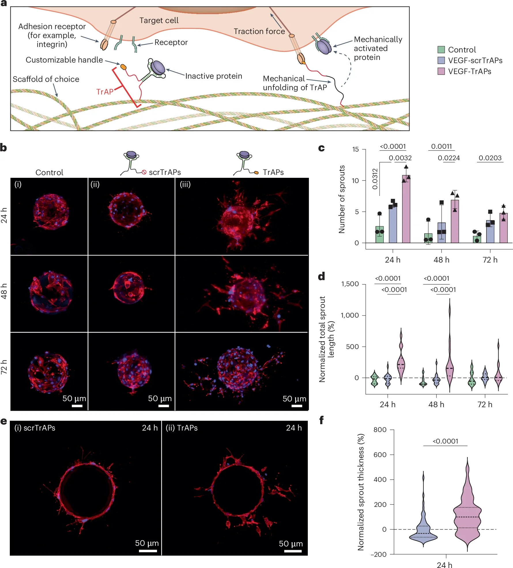
The system uses traction-force-activated payloads, or TrAPs, attached to a scaffold such as collagen. Each contains a folded DNA molecule called an aptamer that binds a particular growth factor and inhibits its activity.
One end anchors the construct to the material. The other carries a short adhesive peptide that gives cells something to grip.
As a cell attaches and pulls, the force unfolds the aptamer. That releases the growth factor near the cell applying the force, linking activation to direct engagement with the scaffold.
Control constructs helped test that mechanism. They retained the growth-factor-binding aptamer but carried a scrambled peptide that could not provide the same cellular grip.
That comparison helped distinguish the effect of holding a protein from the additional effect of allowing cells to release it.
The first experiments focused on vascular endothelial growth factor A, or VEGF-A. It supports blood vessel formation, making it a useful target for testing whether force-triggered release could influence repair.
In cultured endothelial cells, TrAPs preloaded with VEGF-A encouraged more vessel-like sprouts than control materials. After 24 hours, total sprout length per bead was approximately 200% greater than in the control groups.
Sprouts were also about 100% thicker than those exposed to the non-cell-adhesive control constructs. These measurements describe a laboratory sprouting assay, rather than the growth of complete blood vessels in patients.
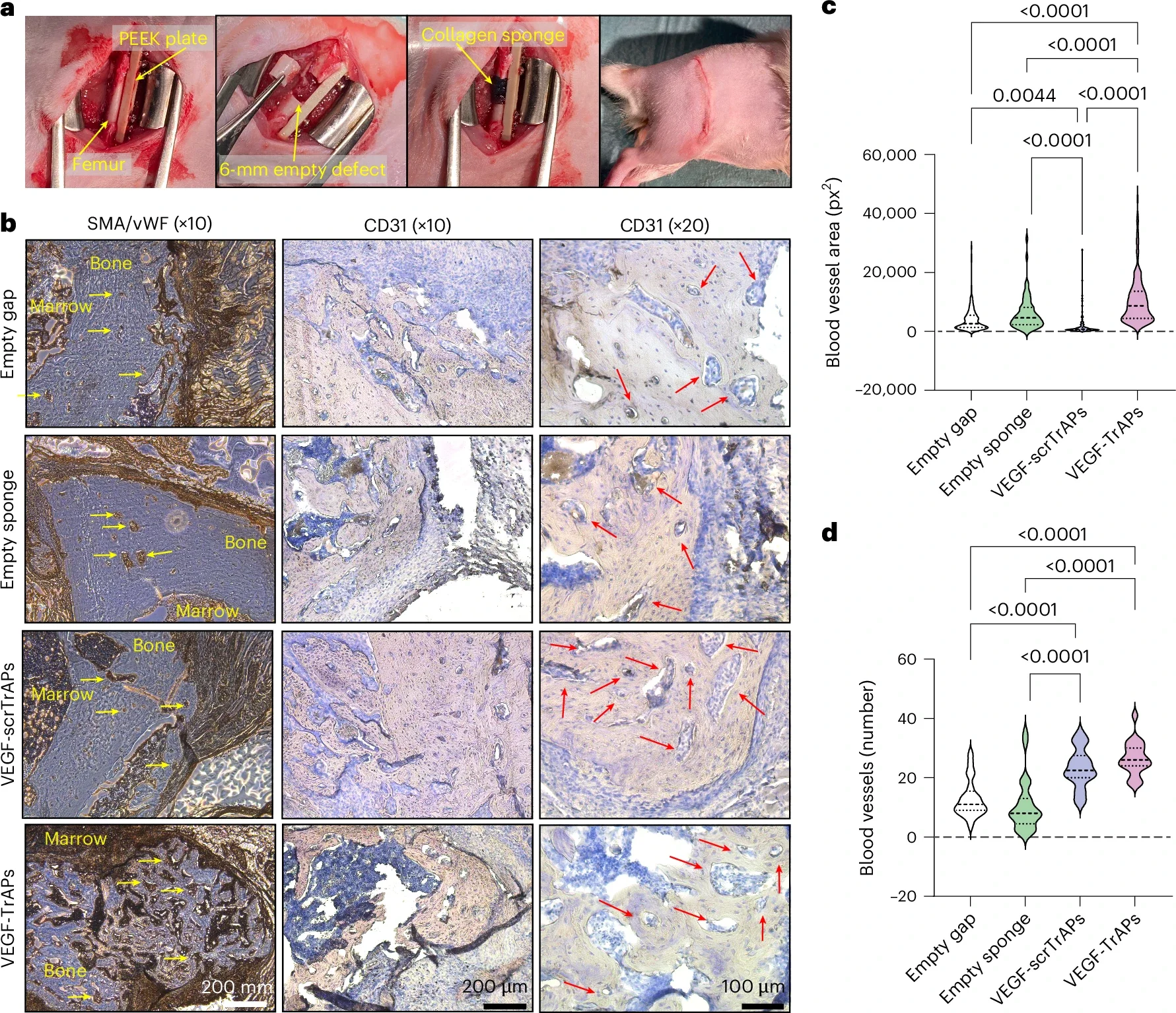
The team then placed VEGF-TrAPs in collagen sponges used in 6-millimeter femoral defects in rats. After three weeks, treated defects contained more blood vessels, with larger vessels than control conditions.
The non-cell-adhesive aptamer controls produced smaller vessels than untreated defects. The researchers interpret this as evidence that those constructs captured naturally available VEGF without releasing it effectively.
No collagen sponges remained at the three-week assessment. The difference between active and inactive constructs nevertheless persisted, supporting a role for cellular activation rather than simple release as the scaffold broke down.
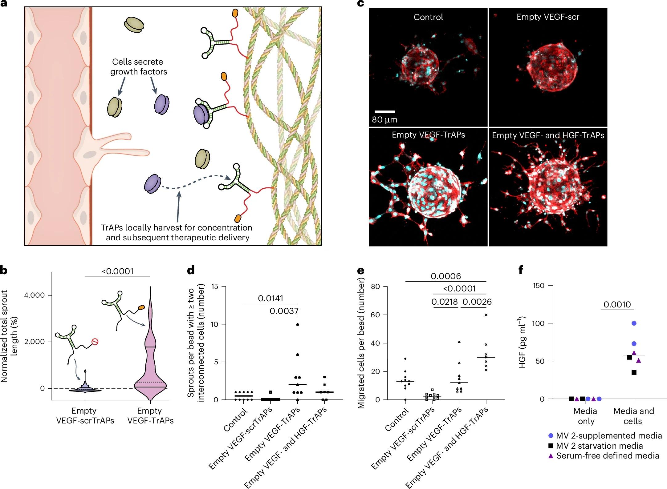
The researchers also tested whether TrAPs could collect proteins already being produced by nearby cells. Empty constructs captured VEGF from human endothelial cells and subsequently supported longer sprouts than inactive aptamer controls.
Another set targeted hepatocyte growth factor, or HGF. Combining the VEGF and HGF systems increased cell migration, suggesting that the material could deliver more than one repair signal.
That flexibility matters because tissue repair involves several coordinating proteins. The study investigated selective capture and release rather than assuming that one growth factor could address every aspect of healing.
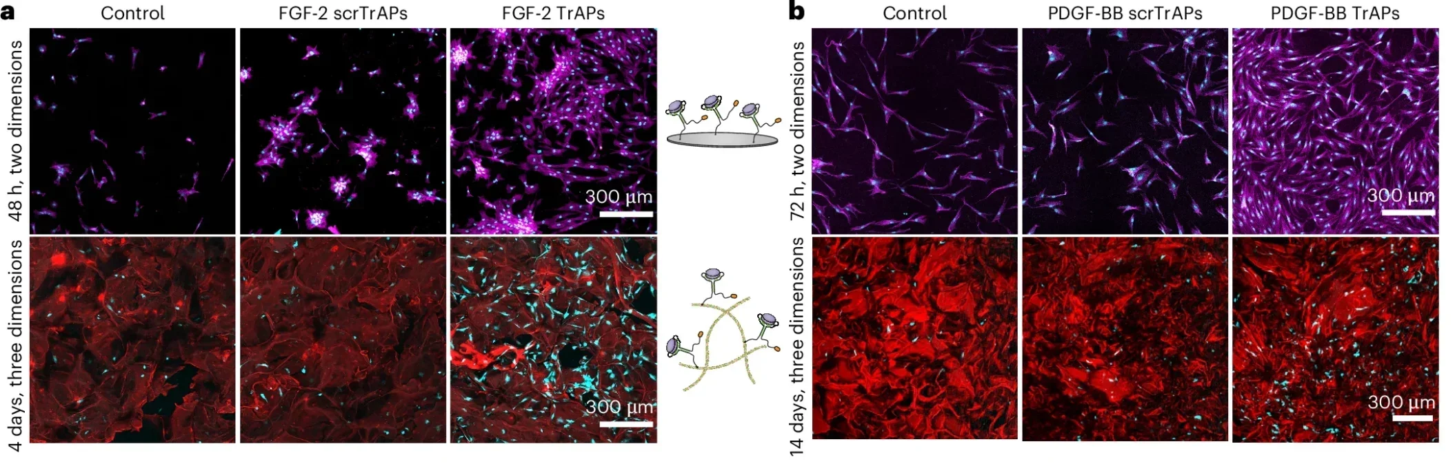
Human platelet lysate provided another source of proteins. This blood-derived preparation contains growth factors released from platelets, which the researchers harvested using different aptamers.
They developed constructs targeting VEGF-A, HGF, fibroblast growth factor-2 and platelet-derived growth factor-BB. Platelet-lysate-loaded TrAPs supported fibroblast growth on flat surfaces and within three-dimensional collagen sponges.
For the human-skin experiments, collagen sponges carried platelet-lysate-loaded constructs targeting FGF-2, PDGF-BB and HGF. After eight days, imaging showed greater overlap between cells and the treated sponge than in control conditions.
Additional staining confirmed migrating keratinocytes at the tissue–sponge interface. These cells form the skin’s outer layer, making their movement relevant to the restoration of a damaged surface.
“What excites me most is that this works in living human skin,” Ho said. The result showed engagement with human tissue, while leaving patient outcomes untested.
The mouse experiments used collagen sponges carrying all four growth-factor-targeting systems, including VEGF-A. They treated 6-millimeter full-thickness skin wounds and assessed changes over time.
After 10 days, the active TrAP group had significantly smaller normalized wound diameters than either control group. Wound-bed thickness showed an upward trend, rather than a confirmed significant improvement.
At that endpoint, the diameter analysis included nine control wounds, 11 inactive-construct wounds and 12 active-construct wounds. Wounds whose sponges fell off were excluded, a limitation relevant to interpreting the comparison.
Early imaging also suggested possible cell infiltration into treated scaffolds. The paper describes that interpretation as unproven, so it cannot establish how much this process contributed to wound closure.
The authors report growth-factor amounts far below those used in several existing delivery approaches. However, comparisons across different proteins, formulations and injuries do not establish equivalent clinical effectiveness.
Capturing proteins from wounds or blood could reduce reliance on manufactured growth factors. Any savings or reduced cold-storage requirements remain possibilities for future development, rather than demonstrated benefits in routine care.
The DNA constructs retained function in wound environments despite their exposure to degrading enzymes. The researchers suggest that attachment to the scaffold helped protect them, although further work must clarify their durability.
No adverse events or animal losses were reported during the study. That finding does not settle questions about human toxicity, immune responses, dosing or long-term tissue remodeling.
Imperial is developing the technology through the Traxion Biotech spinout opportunity. Its clinical path still requires testing, but the experiments establish a useful starting point: cells can trigger local protein delivery while engaging with a repairing tissue.
These studies explore cell-controlled delivery, platelet-derived proteins and the challenges of translating repair signals into treatments.
Biologically Inspired, Cell-Selective Release of Aptamer-Trapped Growth Factors by Traction Forces: The earlier TrAP study demonstrates how cellular pulling can activate aptamer-bound growth factors in laboratory experiments. (Advanced Materials, 2019)
Growth factors and growth factor gene therapies for treating chronic wounds: This review examines delivery approaches, combined growth-factor strategies and barriers to clinical translation. (Bioengineering & Translational Medicine, 2024)
Combination Growth Factor Therapy via Electrostatically Assembled Wound Dressings Improves Diabetic Ulcer Healing In Vivo: An earlier experimental dressing study investigates coordinated delivery of multiple growth factors in diabetic wound models. (Advanced Healthcare Materials, 2015)
Human platelet lysate-cultured adipose-derived stem cell sheets promote angiogenesis and accelerate wound healing via CCL5 modulation: This experimental work explores another use of platelet lysate in supporting cells involved in repair. (Stem Cell Research & Therapy, 2024)
GelMA loaded with platelet lysate promotes skin regeneration and angiogenesis in pressure ulcers by activating STAT3: Researchers examine platelet lysate delivered through a hydrogel in experimental pressure-ulcer repair. (Scientific Reports, 2024)
Research findings are available online in the journal Nature.
The original story “Cell-activated wound dressing releases healing proteins in preclinical tests” is published in The Brighter Side of News.
Like these kind of feel good stories? Get The Brighter Side of News’ newsletter.
The post Cell-activated wound dressing releases healing proteins in preclinical tests appeared first on The Brighter Side of News.


Leave a comment
You must be logged in to post a comment.